Home › Unlabelled › Right Shoulder Anatomy Diagram / File Human Arm Bones Diagram Svg Wikipedia
Right Shoulder Anatomy Diagram / File Human Arm Bones Diagram Svg Wikipedia
Right Shoulder Anatomy Diagram / File Human Arm Bones Diagram Svg Wikipedia. However, more serious injuries, such as complete rotator cuff tears, may require surgical repair. Shoulder pain, instability and, in some cases, a feeling of grinding, locking or catching while moving the shoulder. What does a torn shoulder labrum feel like? The acromioclavicular joint is formed by an articulation between the lateral end of the clavicle and the acromion process of the scapula. The most common symptoms of a torn shoulder labrum are:
This diagram depicts shoulder muscles anatomy diagram. Name this muscle the largest of the shoulder group. The following is an overview of the shoulder muscle anatomy. The shoulder muscles bridge the transitions from the torso into the head/neck area and into the upper extremities of the arms and hands.for that reason, and because of the dexterity of the shoulder joint itself, the musculature of the shoulder is complex, ranging from massive prime mover muscles to finer stabilizer and fixator muscles. The glenohumeral joint is where the ball (humeral head) and the socket (the glenoid) meet.
Shoulder Anatomy Shoulderbuzz from 1pmuda45yr3y2jt8n9364kf9-wpengine.netdna-ssl.com This is the smallest rotator cuff muscle. The upper end of the biceps muscle has two tendons that attach it to bones in the shoulder. The shoulder girdle includes three bones—the scapula, clavicle and humerus. The acromioclavicular joint is where the acromion, part of the shoulder blade (scapula) and the collar bone (clavicle) meet. This is the main muscle that lets you rotate and extend your shoulder. Discuss tha agaonist/antagonist relationship of muscles. Numerous muscles help stabilize the three joints of. Plus, exercises for training them.
Illustration of the shoulder anatomy and labrum.
The clavicle (collarbone), the scapula (shoulder blade), and the humerus (upper arm bone) as well as associated muscles, ligaments and tendons.all together it is made up of four joints as well as the muscles that are responsible for movement in the shoulder attached to the scapula. Back muscles chart 12 photos of the back muscles chart back muscles chart, back muscles diagram and ligaments, back muscles diagram lats, back muscles diagram massage, upper back muscles chart, human muscles, back muscles chart, back muscles diagram and ligaments, back muscles diagram lats, back muscles diagram. This is the smallest rotator cuff muscle. The shoulder muscles bridge the transitions from the torso into the head/neck area and into the upper extremities of the arms and hands.for that reason, and because of the dexterity of the shoulder joint itself, the musculature of the shoulder is complex, ranging from massive prime mover muscles to finer stabilizer and fixator muscles. Learn about these muscles, their origin and insertion points, and their functional anatomy. These muscles form the outer shape of the shoulder and underarm. Sechrest, md narrates an animated tutorial on the basic anatomy of the shoulder. Deltoids anatomy when most people think of the The labrum also serves as the attachment of a major tendon in the shoulder, the biceps tendon. Corey chakarun from shin imaging in california. Name this muscle the largest of the shoulder group. To further reinforce the shoulder, the four muscles of the rotator cuff extend from the scapula and surround the head of the humerus to both rotate the arm and prevent dislocation. There are actually four joints that make up the shoulder.
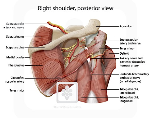
The most common symptoms of a torn shoulder labrum are: Radiology department of the rijnland hospital, leiderdorp and the onze lieve vrouwe. A numeric illustration was then added to show bone anatomy, muscles attachments, ligaments and muscle layers of the rotator cuff. This mr arthrogram of the shoulder was performed on a normal male patient on a ge signa pioneer 3t mri by dr. The following is an overview of the shoulder muscle anatomy.
Shoulder Anatomy New York Ny Handsport Surgery Institute from handsurgeonsnyc.com Back muscles chart 12 photos of the back muscles chart back muscles chart, back muscles diagram and ligaments, back muscles diagram lats, back muscles diagram massage, upper back muscles chart, human muscles, back muscles chart, back muscles diagram and ligaments, back muscles diagram lats, back muscles diagram. Most people with rotator cuff injuries can recover with rest and physical therapy. Anatomy of the shoulder interesting facts about shoulder anatomy. On the anterior side of the shoulder, the coracobrachialis, serratus anterior, pectoralis major, and pectoralis minor muscles work as a group to flex and adduct the scapula and humerus anteriorly toward the sternum. The shoulder girdle includes three bones—the scapula, clavicle and humerus. Learn about these muscles, their origin and insertion points, and their functional anatomy. There are actually four joints that make up the shoulder. The shoulder is a complex combination of bones and joints where many muscles act to provide the widest range of motion of any part of the body.
In this episode of eorthopodtv, orthopaedic surgeon randale c.
The acromioclavicular joint is where the acromion, part of the shoulder blade (scapula) and the collar bone (clavicle) meet. These symptoms may vary depending on the type of labral tear a person has. Corey chakarun from shin imaging in california. The most common symptoms of a torn shoulder labrum are: Sechrest, md narrates an animated tutorial on the basic anatomy of the shoulder. To further reinforce the shoulder, the four muscles of the rotator cuff extend from the scapula and surround the head of the humerus to both rotate the arm and prevent dislocation. Back muscles chart 12 photos of the back muscles chart back muscles chart, back muscles diagram and ligaments, back muscles diagram lats, back muscles diagram massage, upper back muscles chart, human muscles, back muscles chart, back muscles diagram and ligaments, back muscles diagram lats, back muscles diagram. A numeric illustration was then added to show bone anatomy, muscles attachments, ligaments and muscle layers of the rotator cuff. The acromioclavicular joint is formed by an articulation between the lateral end of the clavicle and the acromion process of the scapula. The shoulder is one of the largest and most complex joints in the body. The bones of the shoulder are the humerus (the upper arm bone), the scapula (the shoulder blade), and the clavicle (the collar bone). The upper end of the biceps muscle has two tendons that attach it to bones in the shoulder. Radiology department of the rijnland hospital, leiderdorp and the onze lieve vrouwe.
The long head attaches to the top of the shoulder socket (glenoid). The shoulder has about eight muscles that attach to the scapula, humerus, and clavicle. Editor · aug 6, 2017 ·. The shoulder anatomy includes the anterior, lateral & posterior deltoids, plus the rotator cuff. Discuss tha agaonist/antagonist relationship of muscles.
Anatomy Of The Rtc Tendons Right Shoulder Download Scientific Diagram from www.researchgate.net The glenohumeral joint is where the ball (humeral head) and the socket (the glenoid) meet. Other important bones in the shoulder include: All of the nerves that travel down the arm pass through the axilla (the armpit) just under the shoulder joint and are known as the brachial plexus before dividing into the individual nerves.these nerves carry the signals from the brain to the muscles that move the arm. These symptoms may vary depending on the type of labral tear a person has. In this episode of eorthopodtv, orthopaedic surgeon randale c. On the anterior side of the shoulder, the coracobrachialis, serratus anterior, pectoralis major, and pectoralis minor muscles work as a group to flex and adduct the scapula and humerus anteriorly toward the sternum. There are actually four joints that make up the shoulder. Illustration of the shoulder anatomy and labrum.
The upper end of the biceps muscle has two tendons that attach it to bones in the shoulder. On the anterior side of the shoulder, the coracobrachialis, serratus anterior, pectoralis major, and pectoralis minor muscles work as a group to flex and adduct the scapula and humerus anteriorly toward the sternum. The anatomy of the shoulder. Human anatomy diagram shoulder anatomy shoulder muscles shoulder muscles and chest. The short head attaches to a bump on the shoulder blade called the coracoid process. Numerous muscles help stabilize the three joints of. Name this muscle the largest of the shoulder group. They cover the head of your upper arm bone and attach it to your shoulder blade. Discuss tha agaonist/antagonist relationship of muscles. Back muscles chart 12 photos of the back muscles chart back muscles chart, back muscles diagram and ligaments, back muscles diagram lats, back muscles diagram massage, upper back muscles chart, human muscles, back muscles chart, back muscles diagram and ligaments, back muscles diagram lats, back muscles diagram. Anatomy of the shoulder interesting facts about shoulder anatomy. The labrum is a rim of cartilage that surrounds the socket of the shoulder joint. Rotator cuff injuries are very common, affecting over 3 million people in the united states every year.
The roof of the shoulder is formedby a part of the scapula called the acromion shoulder anatomy diagram. The human shoulder is made up of three bones:


comment 0 comments
more_vert